
Openai/695bc248-c9bc-8008-82d2-be391b6b7b32
This conversation was exported from ChatGPT[1]
gpt-5-2-thinking[edit | edit source]
User: radiographic findings is[edit | edit source]
radiographic findings is consistent with dorsal intercalated segment instability (DISI)ممكن تشرح وتنزل وتجيب صور كثيرة
Assistant: !https://www\.researchgate\.net/profile/Jonny\-Andersson/publication/319937516/figure/fig2/AS%3A540741919559680%4015059…[edit | edit source]
!https://www\.researchgate\.net/profile/Jonny\-Andersson/publication/319937516/figure/fig2/AS%3A540741919559680%401505934328078/Dorsal\-intercalated\-segment\-instability\-DISI\-with\-a\-scapholunate\-angle\-of\-approximately\_Q640\.jpg[2] !https://www\.medical\.theclinics\.com/cms/10\.1016/j\.mcna\.2014\.04\.003/asset/60d34e96\-d732\-48d4\-acd8\-915ab709ac0b/main\.assets/gr8\_lrg\.jpg[3] !https://data\.pacs\.de/sites/default/files/mirrorimages/media\.springernature\.com/full/springer\-static/image/art%253A10\.1007%252Fs13244\-014\-0337\-1/MediaObjects/13244\_2014\_337\_Fig3\_HTML\.gif[4] !https://image\.slidesharecdn\.com/20150914stafflectureupperext\-150913143227\-lva1\-app6891/95/emergency\-radiology\-shoulder\-upper\-extremities\-70\-638\.jpg?cb=1442154947[5]
يا محمد، لما يقول لك السؤال: “radiographic findings consistent with DISI” فهو غالبًا يريد منك “تلتقط” ميل الـ lunate للخلف (dorsiflexion) على lateral wrist radiograph وتثبت ذلك بالأرقام والزوايا الكلاسيكية.
DISI = Dorsal Intercalated Segment Instability[edit | edit source]
- “Intercalated segment” المقصود بها lunate (تُعتبر قطعة “وسطية” بين radius وcarpus).
- في DISI: الـ lunate تميل dorsally (extension/dorsiflexion) لأن التوازن الرباطي اتكسر غالبًا بسبب Scapholunate ligament injury → scaphoid يميل للـ flexion بينما lunate تُسحب للـ extension مع triquetrum/capitate.
المسبب الأشهر:
- Scapholunate ligament injury (خصوصًا بعد FOOSH) وهو المصدر الأكثر كلاسيكية لــ DISI. orthobullets.com[6]
###[edit | edit source]
لازم تكون الصورة true lateral (ده فخ امتحاني: لو الـ lateral غلط الزوايا كلها هتضرب).
- Scapholunate angle (SLA) مرتفع
- الطبيعي تقريبًا 30° إلى 60°.
- في DISI: غالبًا SLA أكبر من 70° (وهي عبارة امتحانية صريحة جدًا). orthobullets.com[7]
- Radiolunate angle (RLA) مرتفع
- في DISI يحدث dorsal tilt of the lunate ويظهر كـ RLA أكبر من 10°. Radiopaedia[8]
- Capitolunate angle (أو Lunocapitate angle) مرتفع
- كثير من المراجع تذكر Capitolunate angle أكبر من 30° كداعم قوي للـ DISI. PMC[9]
1. Scapholunate gap widening[edit | edit source]
- “Terry Thomas sign” (زيادة المسافة بين scaphoid وlunate).
- أحيانًا تحتاج stress views مثل clenched fist view لإظهار الـ widening إذا كانت الإصابة ديناميكية. pubs.rsna.org[10]
- Scaphoid ring sign
- نتيجة scaphoid flexion/rotary subluxation (حلقة/دائرة واضحة في scaphoid على PA).
- على lateral: - ارسم محور طويل لـ scaphoid ومحور لـ lunate → ده يعطيك Scapholunate angle. - محور radius مع محور lunate → Radiolunate angle. - محور capitate مع محور lunate → Capitolunate angle. الفكرة موجودة بصريًّا في الصور المرفقة في الأعلى (خصوصًا الرسوم التعليمية).[edit | edit source]
- ليست كل زيادة في SLA = DISI إذا كانت اللقطة ليست true lateral أو كان المعصم في وضع غير neutral. PMC[11][edit | edit source]
- DISI قد تكون dynamic: PA طبيعي تقريبًا لكن تظهر بالـ clenched fist أو ulnar deviation stress. pubs.rsna.org[12]
- فرّق بين DISI و VISI: - DISI عادة مع Scapholunate ligament injury - VISI عادة مع Lunotriquetral ligament injury (وهنا lunate تميل volarly). PMC[13]
الـ DISI المزمنة غالبًا تمشي في مسار SLAC wrist (Scapholunate Advanced Collapse)، ودي فكرة امتحانية شائعة جدًا: إصابة SL → DISI → تغيّرات تنكسية تدريجية. PMC[14][edit | edit source]
- لو الأشعة مش واضحة والشك عالي: - اعمل stress views (clenched fist). pubs.rsna.org[15] - وMRI / MR arthrogram لتقييم Scapholunate ligament خصوصًا في الإصابات الحادة/قبل الجراحة. pubs.rsna.org[16][edit | edit source]
إذا تحب، ابعتلي صورة السؤال/الأشعة اللي عندك (خصوصًا الـ lateral)، وأنا أمشي معك خطوة بخطوة: هل هي true lateral؟ وهل الزوايا داخلة في DISI ولا في perilunate/lunate dislocation (لأن دي برضه أسئلة شائعة وتتلخبط بسهولة).
gpt-5-2[edit | edit source]
User: ممكن تعيد بالانجليزي[edit | edit source]
ممكن تعيد بالانجليزي
Assistant: !https://prod\-images\-static\.radiopaedia\.org/images/30733038/986af30d0ca20e140b04cf5487c016\big\gallery\.jpeghttps:/…[edit | edit source]
!https://prod\-images\-static\.radiopaedia\.org/images/30733038/986af30d0ca20e140b04cf5487c016\_big\_gallery\.jpeg[17] !https://www\.researchgate\.net/profile/Ahmed\-Elsaftawy/publication/256452003/figure/fig5/AS%3A314936254320644%401452098062635/n\-case\-of\-rotary\-subluxation\-of\-the\-scaphoid\-scapholunate\-angle\-60\-lunate\-capitate\_Q320\.jpg[18] !https://upload\.orthobullets\.com/topic/6041/images/gw275h550\.\.jpg[19] 5
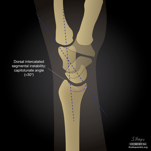
Radiographic Findings Consistent with DISI (Dorsal Intercalated Segment Instability)[edit | edit source]
1) What does DISI mean?[edit | edit source]
DISI is a pattern of carpal instability in which the lunate tilts dorsally (extends) on the lateral wrist radiograph. It occurs most commonly after Scapholunate ligament injury. Mechanism in brief:
- Scaphoid loses its stabilizing link to the lunate → scaphoid collapses into flexion
- Lunate, pulled by the triquetrum and capitate, rotates into extension (dorsiflexion)
2) Key Radiographic Findings (High-yield for exams)[edit | edit source]
A) Lateral view (the most important view)[edit | edit source]
- The diagnosis of DISI is primarily made on a true lateral wrist radiograph.
- Dorsal tilt of the lunate
- The lunate’s distal articular surface faces dorsally
- Often described as the lunate “cup” opening posteriorly
- Increased Scapholunate Angle (SLA)
- Normal: approximately 30°–60°
- DISI: typically greater than 70°
- This is the single most quoted numeric criterion in exams
- Increased Radiolunate Angle (RLA)
- Normal: near neutral
- DISI: greater than 10°, reflecting dorsal lunate tilt
- Increased Capitolunate (Lunocapitate) Angle
- Normal: less than 30°
- DISI: greater than 30°, indicating loss of collinearity between lunate and capitate
- Classic exam phrase:
- “DISI on lateral view is characterized by dorsal lunate tilt with an increased scapholunate angle, usually greater than 70 degrees.”
B) PA (posteroanterior) view – supportive findings[edit | edit source]
PA views help identify the cause, not the alignment itself.
- Scapholunate gap widening
- Increased distance between scaphoid and lunate
- Known as the “Terry Thomas sign”
- Scaphoid ring sign
- Seen due to scaphoid flexion and rotary subluxation
- Dynamic instability
- Early DISI may have a normal PA view
- Widening appears only on stress views such as: - Clenched fist view - Ulnar deviation stress view
3) How to Measure (Practical Exam Technique)[edit | edit source]
On a true lateral radiograph:
- Draw the long axis of the scaphoid
- Draw the axis of the lunate → angle between them = Scapholunate angle
- Draw the radius axis and lunate axis → Radiolunate angle
- Draw the capitate axis and lunate axis → Capitolunate angle
If the lateral is not perfectly true, angles are unreliable (important pitfall).
4) Common Pitfalls (Frequently Tested)[edit | edit source]
- False DISI can be diagnosed if the lateral view is rotated or not neutral
- DISI vs VISI: - DISI → dorsal lunate tilt → Scapholunate ligament injury - VISI → volar lunate tilt → Lunotriquetral ligament injury
- Early DISI can be dynamic, visible only with stress imaging
5) Why DISI Matters Clinically (Exam Progression)[edit | edit source]
Untreated or chronic DISI commonly progresses to:
- SLAC wrist (Scapholunate Advanced Collapse)
- Predictable pattern of degenerative arthritis
This progression is a very common MCQ concept.
6) If X-rays Are Inconclusive[edit | edit source]
Next diagnostic steps may include:
- Stress radiographs
- MRI or MR arthrogram to assess the Scapholunate ligament
- Wrist arthroscopy (gold standard in equivocal cases)
One-line exam summary[edit | edit source]
- DISI is diagnosed on a lateral wrist radiograph by dorsal tilting of the lunate with an increased scapholunate angle (usually greater than 70°), most commonly due to scapholunate ligament injury.
If you want, send me a specific wrist X-ray and I’ll walk you through it step-by-step exactly as in a viva or OSCE scenario.
References[edit | edit source]
- ↑ "DISI شرح ونتائج إشعاعية". ChatGPT. Retrieved 2026-01-06.
- ↑ "https://www\.researchgate\.net/profile/Jonny\-Andersson/publication/319937516/figure/fig2/AS%3A540741919559680%401505934328078/Dorsal\-intercalated\-segment\-instability\-DISI\-with\-a\-scapholunate\-angle\-of\-approximately\_Q640\.jpg". tse1.mm.bing.net. Retrieved 2026-01-06.
{{cite web}}: External link in|title= - ↑ "https://www\.medical\.theclinics\.com/cms/10\.1016/j\.mcna\.2014\.04\.003/asset/60d34e96\-d732\-48d4\-acd8\-915ab709ac0b/main\.assets/gr8\_lrg\.jpg". tse3.mm.bing.net. Retrieved 2026-01-06.
{{cite web}}: External link in|title= - ↑ "https://data\.pacs\.de/sites/default/files/mirrorimages/media\.springernature\.com/full/springer\-static/image/art%253A10\.1007%252Fs13244\-014\-0337\-1/MediaObjects/13244\_2014\_337\_Fig3\_HTML\.gif". tse2.mm.bing.net. Retrieved 2026-01-06.
{{cite web}}: External link in|title= - ↑ "https://image\.slidesharecdn\.com/20150914stafflectureupperext\-150913143227\-lva1\-app6891/95/emergency\-radiology\-shoulder\-upper\-extremities\-70\-638\.jpg?cb=1442154947". tse3.mm.bing.net. Retrieved 2026-01-06.
{{cite web}}: External link in|title= - ↑ "orthobullets.com". orthobullets.com. Retrieved 2026-01-06.
- ↑ "orthobullets.com". orthobullets.com. Retrieved 2026-01-06.
- ↑ "Radiopaedia". radiopaedia.org. Retrieved 2026-01-06.
- ↑ "PMC". pmc.ncbi.nlm.nih.gov. Retrieved 2026-01-06.
- ↑ "pubs.rsna.org". pubs.rsna.org. Retrieved 2026-01-06.
- ↑ "PMC". pmc.ncbi.nlm.nih.gov. Retrieved 2026-01-06.
- ↑ "pubs.rsna.org". pubs.rsna.org. Retrieved 2026-01-06.
- ↑ "PMC". pmc.ncbi.nlm.nih.gov. Retrieved 2026-01-06.
- ↑ "PMC". pmc.ncbi.nlm.nih.gov. Retrieved 2026-01-06.
- ↑ "pubs.rsna.org". pubs.rsna.org. Retrieved 2026-01-06.
- ↑ "pubs.rsna.org". pubs.rsna.org. Retrieved 2026-01-06.
- ↑ "https://prod\-images\-static\.radiopaedia\.org/images/30733038/986af30d0ca20e140b04cf5487c016\_big\_gallery\.jpeg". prod-images-static.radiopaedia.org. Retrieved 2026-01-06.
{{cite web}}: External link in|title= - ↑ "https://www\.researchgate\.net/profile/Ahmed\-Elsaftawy/publication/256452003/figure/fig5/AS%3A314936254320644%401452098062635/n\-case\-of\-rotary\-subluxation\-of\-the\-scaphoid\-scapholunate\-angle\-60\-lunate\-capitate\_Q320\.jpg". images.openai.com. Retrieved 2026-01-06.
{{cite web}}: External link in|title= - ↑ "https://upload\.orthobullets\.com/topic/6041/images/gw275h550\.\.jpg". upload.orthobullets.com. Retrieved 2026-01-06.
{{cite web}}: External link in|title=